
Hip health plays a pivotal role in our daily lives, influencing everything from walking to sitting comfortably. When hip problems arise, they can significantly impact mobility and quality of life. Fortunately, MRI pelvis scans offer a detailed look into hip health, providing valuable insights that help diagnose and treat various hip conditions effectively. This article delves into the world of MRI pelvis scans, exploring how they work, what they can reveal about hip health, and why they are instrumental in modern healthcare.
Understanding MRI Pelvis Scans
What is an MRI Pelvis Scan?
MRI, or Magnetic Resonance Imaging, is a sophisticated medical imaging technique that uses powerful magnets and radio waves to generate detailed images of the body’s internal structures. Specifically, an MRI pelvis scan focuses on capturing images of the hip joint and surrounding areas within the pelvis. Unlike X-rays or CT scans, MRI does not use ionizing radiation, making it a safer option for repeated imaging.
Advantages of MRI Pelvis Scans
One of the primary benefits of MRI pelvis scans is their ability to produce high-resolution images of bones, cartilage, muscles, and soft tissues in and around the hip joint. This level of detail is crucial for diagnosing a wide range of hip conditions, from fractures and arthritis to soft tissue injuries like labral tears. Additionally, MRI provides multi-planar imaging capabilities, allowing healthcare providers to view the hip joint from different angles for a comprehensive assessment.
Anatomy of the Hip and Pelvis
Key Structures of the Hip
The hip joint is a complex structure consisting of several key components:
- Bones: The femur (thigh bone) articulates with the pelvis to form the hip joint. The acetabulum, a socket in the pelvic bone, holds the ball-shaped head of the femur.
- Cartilage: Articular cartilage covers the surfaces of the bones where they meet at the hip joint, providing smooth movement.
- Muscles and Ligaments: Various muscles, including the hip flexors, abductors, adductors, and gluteal muscles, surround the hip joint, providing stability and movement. Ligaments such as the iliofemoral, pubofemoral, and ischiofemoral ligaments help reinforce the joint.
Common Hip Problems and Conditions
Numerous conditions can affect the hip joint and surrounding structures, including:
- Osteoarthritis: Degenerative joint disease characterized by the breakdown of cartilage and bone within the hip joint.
- Labral Tears: Tears in the labrum, a ring of cartilage that surrounds the acetabulum, often caused by repetitive motions or trauma.
- Femoroacetabular Impingement (FAI): A condition where there is abnormal contact between the ball and socket of the hip joint, leading to pain and restricted movement.
- Hip Fractures and Stress Injuries: Fractures of the femur or pelvic bones, which can occur due to trauma or overuse injuries.
When is an MRI Pelvis Scan Needed?
Diagnostic Indications
Healthcare providers may recommend an MRI pelvis scan under various circumstances, including:
- Persistent hip pain or discomfort that does not improve with rest or over-the-counter medications.
- Difficulty walking, standing, or performing daily activities due to hip stiffness or instability.
- Suspected soft tissue injuries or structural abnormalities identified during physical examination.
Conditions Diagnosed by MRI
MRI pelvis scans are instrumental in diagnosing a wide range of hip conditions, including:
- Soft Tissue Injuries: Such as labral tears, tendonitis, or bursitis.
- Structural Abnormalities: Including hip dysplasia or congenital abnormalities.
- Degenerative Changes: Such as osteoarthritis or avascular necrosis (bone tissue death).
- Pre- and Post-Surgical Evaluations: Providing detailed images for surgical planning and monitoring post-operative healing.
The MRI Pelvis Scan Procedure
Preparation for the Scan
Before undergoing an MRI pelvis scan, patients may be instructed to:
- Remove jewelry, clothing with metal components, and other metallic objects.
- Inform the technologist of any medical devices or implants, such as pacemakers or joint replacements.
- Follow fasting instructions if contrast dye is required for the scan.
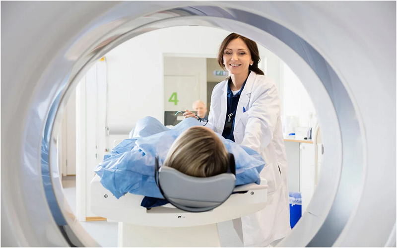
During the Scan
During the MRI pelvis scan:
- Patients lie comfortably on a table that slides into the MRI machine.
- Coils may be placed around the hip area to enhance image quality.
- The MRI machine generates loud tapping or knocking sounds as it creates images, and patients may be provided with earplugs or headphones for comfort.
Post-Scan Process
After the scan:
- Images are reviewed and analyzed by a radiologist, who prepares a detailed report for the referring healthcare provider.
- Patients receive the results of their MRI pelvis scan during a follow-up appointment, where further treatment plans or recommendations may be discussed based on the findings.
Insights Provided by MRI Pelvis Scans
Diagnosing Hip Pathologies
For instance, MRI pelvis scans can accurately diagnose conditions like osteoarthritis by visualizing joint space narrowing and the presence of osteophytes (bone spurs). This information is crucial for determining the appropriate treatment approach, whether it involves medication, physical therapy, or joint replacement surgery.
Assessing Soft Tissue Injuries
Consider a scenario where an athlete experiences persistent hip pain. An MRI pelvis scan can reveal a labral tear or inflammation of the hip tendons, providing insights into the severity of the injury and guiding rehabilitation efforts.
Planning Surgical Interventions
In cases requiring surgical intervention, such as FAI correction or labral repair, MRI pelvis scans offer precise imaging for surgical planning. Surgeons can visualize the affected structures in detail before performing procedures, leading to more successful outcomes and faster recovery times.
Future Trends in MRI Technology
Advancements in MRI Imaging
Continuous advancements in MRI technology include improvements in image resolution and scan speed. These developments enhance diagnostic accuracy and patient comfort during MRI pelvis scans, making them more accessible and efficient in clinical settings.
AI and Image Analysis
The integration of artificial intelligence (AI) in MRI image analysis holds promise for enhancing diagnostic capabilities. AI algorithms can assist radiologists in interpreting MRI pelvis scans more efficiently, leading to faster turnaround times for results and improved patient care outcomes.
Conclusion
MRI pelvis scans are indispensable tools for evaluating hip health and diagnosing a wide range of conditions affecting the hip joint and surrounding structures. By providing detailed images without the use of radiation, MRI technology supports healthcare providers in developing personalized treatment plans tailored to each patient’s needs.
For comprehensive MRI pelvis scans and expert diagnostic imaging services, visit Upright MRI of Deerfield. Our dedicated team is committed to delivering high-quality care and accurate diagnostic assessments to help you achieve optimal hip health. Contact us today to schedule your appointment and take proactive steps toward managing your hip condition effectively.
By understanding what MRI pelvis scans can uncover about hip health, you empower yourself to make informed healthcare decisions and prioritize your well-being. Don’t delay—take charge of your hip health journey today for a more active and pain-free tomorrow!